
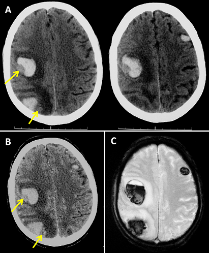
Simultaneous bleeding of multiple brain metastases from pulmonary carcinoma
Ali Akhaddar, Hatim Belfquih
PAMJ-CM. 2020; 3:140. Published 28 Jul 2020 | doi:10.11604/pamj-cm.2020.3.140.24556
Corresponding author
Ali Akhaddar, Department of Neurosurgery, Avicenne Military Hospital of Marrakech, Marrakech, Morocco (akhaddar@hotmail.fr)
This image
| Articles published in PAMJ-CM are Open Access and distributed under the terms of the Creative Commons Attribution 4.0 International (CC BY 4.0). |  |

eISSN: 2707-2797
The PAMJ Clinical Medicine (ISSN: 2707-2797) is a subsidiary of the Pan African Medical Journal. The contents of this journal is intended exclusively for professionals in the medical, paramedical and public health and other health sectors.
Currently tracked by: DOAJ, AIM, Google Scholar, AJOL, EBSCO, Scopus, Embase, IC, HINARI, Global Health, PubMed Central, PubMed/Medline, ESCI
Physical address: Kenya: 3rd Floor, Park Suite Building, Parkland Road, Nairobi. PoBox 38583-00100, tel: +254 (0)20-520-4356 | Cameroon: Immeuble TechnoPark Essos, Yaounde, PoBox: 10020 Yaounde, tel: +237 (0)24-309-5880

About PAMJ - Manuscript Hut™
The Manuscript Hut is a product of the PAMJ Center for Public health Research and Information.
Kenya: 3rd Floor, Park Suite Building, Parkland Road, Nairobi. PoBox 38583-00100, tel: +254 (0)20-520-4356
Cameroon: Immeuble TechnoPark Essos, Yaounde, PoBox: 10020 Yaounde, tel: +237 (0)24-309-5880
Copyright © - Pan African Medical Journal - CEPHRI. 2026
Haraka Publishing Platform - (MMS V.2.5). Release date Jan 2018 - Customized for PAMJ - Clinical Medicine
services@panafrican-med-journal.com
 |